Wednesday, March 29, 2017

Brain Dissection Lab


2. Cerebrum- takes information and processes it
cerebellum- motor control and muscle movement
brain stem- connects to spinal cord, breathing, digestion
meninges- separates blood and brain

3. Insulation around the neuron. Allows faster communication

4.


5.  Thalamus- relays sensory information, feels pain
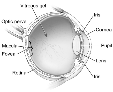
Optic Nerve- connects the brain to the eye
Medulla Oblongata- controls heart and lungs
Pons- links the medulla oblongata to the thalamus
Midbrain- vision, hearing, and motor control
Corpus Collusum- joins the two hemispheres of the brain
Hypothalamus- controls the autonomic nervous system


6.


Relate and Review:

This lab was very interesting. I'm getting better at labs in terms of being able to find things easier and also not being grossed out. I think the most interesting part was seeing the corpus callosum separating the two distinct hemispheres of the brain. Doing a lab is different than reading a textbook or watching a vodcast because it isn't color coded and labeled. This leaves a lot of grey area for error, however once you focus on what you're looking for you're able to find it.


Friday, March 17, 2017

Unit 6 Reflextion

This unit was primarily about our brain and nervous system. We learned how the brain is separated into different sections, which control different actions and different parts of the body. We learned how a reflex is when sensory information travels down a synapse to the brain and the brain quickly responds. The brain takes information from our five senses; sight, smell, hear, taste, and touch  We us our eyes to see, nose to smell, ears to hear, mouth to taste, and our whole body to touch. In the brain dissection, we could clearly see how the brain is split in half. The eye dissection seemed complicated at times but i thought it was interesting and helpful. It's funny comparing that dissection to the simplicity of the Hana the Banana dissection. Some weaknesses i have in this unit would be remembering all the parts to the eye, and not getting my brain parts mixed up. A strength would be the five senses. For my New Years goals I have just started lifting.


Reflex Lab Analysis

1. For the Reflex Lab, we conducted experiments to further understand our reflexes. Reflexes, a mechanism to protect our body, is when sensory information travels down a synapse to the brain and the brain responds quickly. 

2. I) In experiment 1 the photo pupillary reflex contracts the smooth muscle of the iris to control the size of the pupil. When we cover our eyes to make it dark, our pupil expands in an attempt to allow more light in. When we are exposed back to the light our pupil then contracts to limit the amount of light getting in. Humans evolved this trait to increase vision in the dark.
II) The patellar reflex causes the knee to jerk in a monosynaptic reflex. It occurs so fast because there is only one synapse in the circuit needed to complete the reflex. Although after the squares were completed, the knee did not jerk because the muscles are tired and cannot react as fast. Humans evolved this reflex in order to react to danger immediately. 
III) The blink reflex is something humans developed to keep things out of our eyes. The cotton ball, or anything coming at our eyes, causes us to blink without thinking to protect our eyes.
IV) For the fourth experiment we used the Plantar Reflex. When we drag the pen on our foot, our toes are supposed to curl. However, people suffering from MS and Newborns spread their toes instead because their nervous system isn't myelinated, resulting in Babinski's sign. 
V) The last test doesn't measure a reflex, but rather your hand-eye coordination. Mine and John's were very similar, with my average time being 0.16 seconds and his being 0.18 seconds. A possible mistake we could have made in this experiment could have been holder the meter stick closer or further away from the hand, or perhaps miscalculation. 

3. Texting significantly slowed down our reaction time. I went from an average of 0.16 seconds without texting to 0.30 seconds with seconds, nearly doubling my time! This gives us significant evidence to prove that texting while driving is very dangerous. 


Monday, March 6, 2017

Sheep Eye Dissection Analysis

Light first travels through the cornea and then passes through the pupil, which is controlled by the iris(colored part of eye). Then the lens bends the light to the optic nerve, which is connected to the brain (left eye=right brain, right eye=left brain). The sclera is the white part. The vitrous humor is the liquid that fills the eye. Fatty tissue surrounds the sclera and protects the eye. Choroid is the second layer of the eye, and is colored black. The cilary body consists of muscles, which control and shape the lens. The tapetum lucidum is found in nocturnal animals. It reflects light and increase vision in the dark. The suspensory ligament attaches the lens to the optic nerve. Aqueous Humor is the rest of liquid that fills the eye.

Image result for eye diagram