Monday, March 6, 2017
Sheep Eye Dissection Analysis
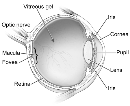
Light first travels through the cornea and then passes through the pupil, which is controlled by the iris(colored part of eye). Then the lens bends the light to the optic nerve, which is connected to the brain (left eye=right brain, right eye=left brain). The sclera is the white part. The vitrous humor is the liquid that fills the eye. Fatty tissue surrounds the sclera and protects the eye. Choroid is the second layer of the eye, and is colored black. The cilary body consists of muscles, which control and shape the lens. The tapetum lucidum is found in nocturnal animals. It reflects light and increase vision in the dark. The suspensory ligament attaches the lens to the optic nerve. Aqueous Humor is the rest of liquid that fills the eye.
Subscribe to:
Post Comments (Atom)
No comments:
Post a Comment